
Fig. 5.
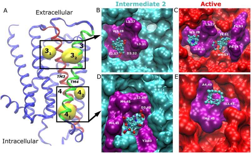
(A) Allosteric sites 3 and 4 are located in the lipid-exposed pockets formed by the TM3 (red)-TM4 (green) extracellular and intracellular domains, respectively. The receptor is rotated from Fig. 2 by 180° along the main axis of the TM bundle for better view of the two sites. The key residues that interact with bound probes (ball-and-sticks) are labeled and highlighted in purple for Site 3 in (B) intermediate 2 and (C) active conformers, and similarly for Site 4 in (D)-(E).