
Fig. 2.
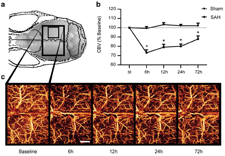
Cortical perfusion is impaired globally early after SAH. a Changes in perfused CBV were tracked in mice for several days following SAH or sham surgery using OMAG. The scan area of the dorsal cortex was 5× 7.5 mm (large box). The smaller box indicates the scan area for all subsequent studies. b Cortical perfusion was reduced in SAH animals as early as 6 h after SAH and persisting for at least 72 h. c Representative OMAG images of SAH mouse showing changes in perfused CBV over time. Decreased intensity corresponds to a reduction in perfused CBV. Values are mean±SEM. SAH (n=10), sham (n=5), *P<0.01. Scale bar= 1 mm