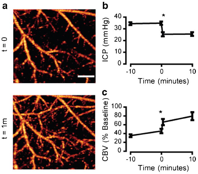
Fig. 3.

Elevated ICP contributes to the early decrease in cortical perfusion after SAH. a Representative OMAG scans of the cortex within the MCA territory 1 h after SAH. A ventricular cannula was placed on the contralateral side and opened to drain CSF, leading to an immediate improvement of cortical perfusion. b ICP measurements 1 h post-SAH. Cannula was opened to allow CSF drainage at time=0. n=5, *P<0.05. c Quantification of perfused CBV 1 h post-SAH. Cannula was opened at time=0. n=5, *P<0.05. Scale bar=500 μm
