Fig. 7.

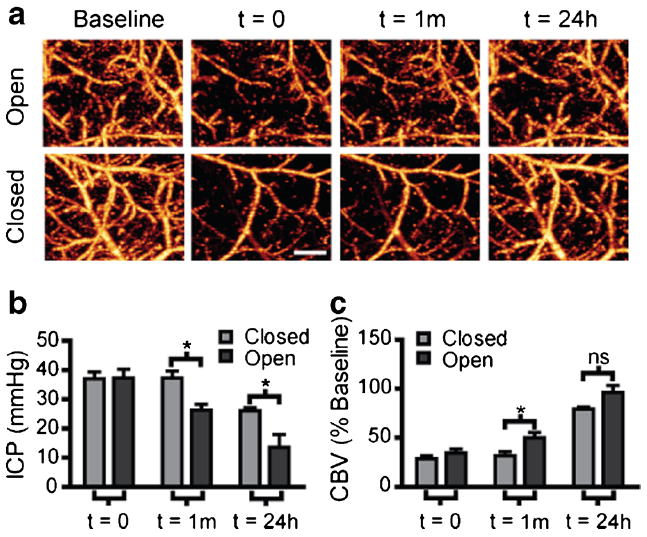
CSF drainage without tPA does not improve cortical perfusion at 24 h. a Representative OMAG images showing changes in perfused CBV. A ventricular cannula was placed on the contralateral side and opened 1 h after SAH then allowed to drain for 24 h. Scans were taken at baseline, 1 h post-SAH before cannula manipulation (time=0), immediately after cannula manipulation (time=1 m), and 24 h later (time=24 h). b Quantification of ICP 1 h post-SAH before cannula manipulation (time=0), immediately after cannula manipulation (time=1 m), and 24 h later (time= 24 h). Open cannula reduced ICP at both the 1 h and 24 h time points after SAH. Closed (n=4), open (n=4), *P<0.05. c Quantification of perfused CBV changes expressed as percent of baseline. Open cannula increased perfused CBV over closed group at 1 h but not 24 h after SAH. Closed (n=6), open (n=8), *=P<0.05, ns no significance. Scale bar=500 μm
