Figure 2.

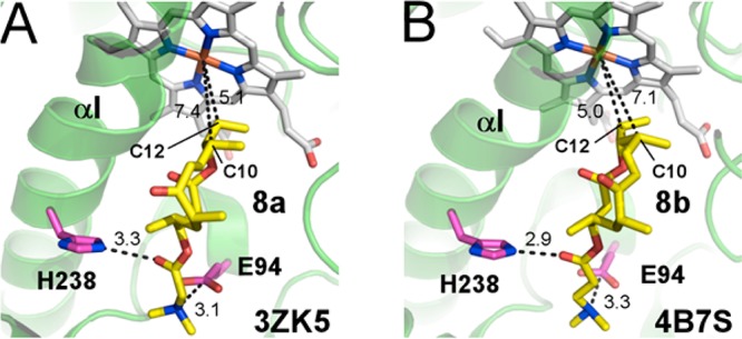
Active site of PikCD50N with 8a (A) and 8b (B) bound. Substrates are in yellow, heme in gray, and amino acid residues in pink sticks; fragments of protein structure are shown as green ribbon. Distances are in angstroms.

Active site of PikCD50N with 8a (A) and 8b (B) bound. Substrates are in yellow, heme in gray, and amino acid residues in pink sticks; fragments of protein structure are shown as green ribbon. Distances are in angstroms.