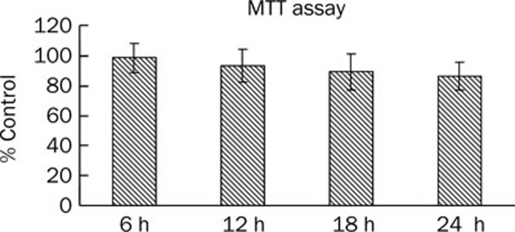
Figure 1.
Effect of ox-LDL on cell viability in HUVECs. HUVECs were seeded in 96-well plates at 103 cells/well and allowed to adhere for 8 h. The subconfluent cells were then exposed to medium containing 40 μg/mL ox-LDL. MTT assay was performed at 0, 6, 12, and 24 h time point. Each experiment was performed in triplicate and bars represent mean±SD. n=3.

