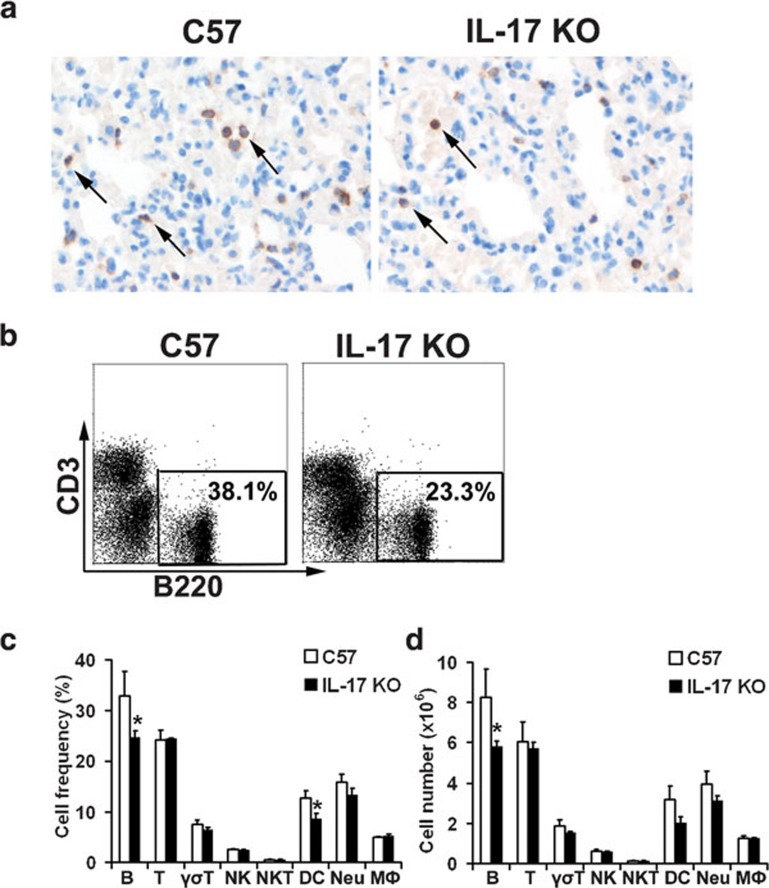
Figure 3.
The total number and frequency of B cells were significantly reduced in the lung tissue of IL-17 KO mice upon virus stimulation. (a) Immunohistochemical staining of B220+ B cells in the lungs collected at 5 dpi. Sections are representative of four mice in each group. Images are at magnification ×400. (b) Flow cytometric analysis of B-cell frequencies among immune cells in the lung 48 h post-stimulation with inactivated virus. Numbers indicate the percentage of B220+ cells. Figures are representative of three mice in each group. (c, d) Cellularity analysis of B cells (B220+), T cells (CD3+), γσT cells (γσTCR+), NK cells (NK1.1+CD3-), NKT cells (NK1.1+CD3+), DCs (CD11c+), neutrophils (Gr1+CD11b+) and macrophages (Gr1−CD11b+) in the lungs 48 h post-stimulation with inactivated virus (n=3 in each group). *P<0.05.DC, dendritic cell; dpi, days post-infection; IL, interleukin; KO, knockout; NK, natural killer; NKT, natural killer T.

