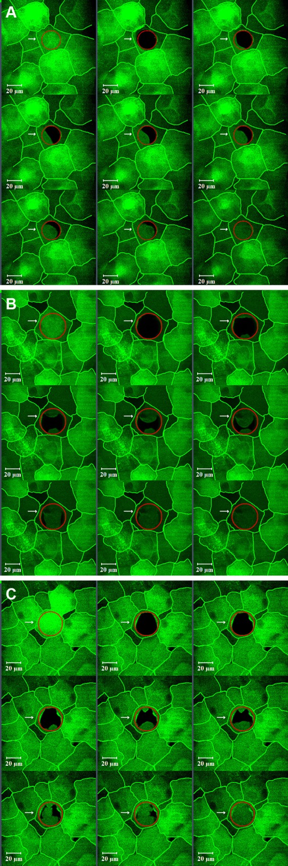
Figure 1.

Different fluorescence recovery patterns observed in corneal epithelial cells. Recovery was seen moving from a either a single direction, typically as was observed in superficial cells from VDR−/− mice (A), or from two (B) or more (C) neighboring cells. Cell borders have been highlighted.
