Figure 4.

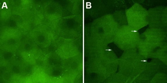
Confocal images obtained from corneal surface of VDR+/+ (A) and VDR−/− mice (B). Superficial squamous cells of VDR−/− mice partly abutted to their neighboring cells. Arrows indicate spaces between cells.

Confocal images obtained from corneal surface of VDR+/+ (A) and VDR−/− mice (B). Superficial squamous cells of VDR−/− mice partly abutted to their neighboring cells. Arrows indicate spaces between cells.