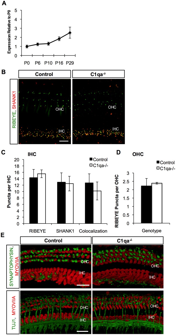
Figure 2. C1qa−/− mice have no alteration in synaptic refinement.
A) Whole cochlea tissue was isolated from mice during ages of synaptic refinement and maturation and C1qa mRNA expression was analyzed. n = 3–5 mice per age group. Error bars show standard error of the mean. B) Double labeling fluorescent images showing RIBEYE (green) and SHANK1 (red) puncta to mark presynaptic and postsynaptic ribbons, respectively in mice P29 of age in both Control and C1qa−/− mice. Scale bar 10 um. C) and D) Quantification of RIBEYE and/or SHANK1 puncta beneath IHCs and OHCs of Control wild type and C1qa−/− mice, respectively, showed no significant differences between the two groups. p = 0.27 for RIBEYE, p = 0.8 for SHANK, and p = 0.22 for colocalized synaptic puncta in IHCs and p = 0.517 for RIBEYE in OHCs. E) Double labeling fluorescent images showing SYNPATOPHYSIN (green) to mark efferent synapses or TUJ1 (green) for efferent and afferent fibers and MYOVIIa (red) to mark IHCs and OHCs in Control and C1qa−/− mice at P29 of age. Scale bar 10 um.

