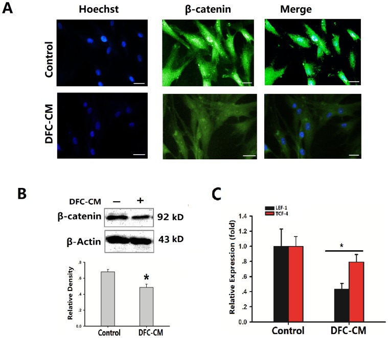
Figure 3. DFC-CM condition effect Wnt/β-catenin signaling pathway in ADSCs.
(A) Immunocytochemical staining showed that ADSCs cultured in basic medium or in DFC-CM condition expressed β-catenin. Scale bar represents 100 µm. (B) Beta-Catenin levels were examined by Western blot analysis and scanning densitometer. Beta-actin was used as internal control. (C). LEF-1and TCF-4 mRNA were subjected to real time-PCR analysis after cells were cultured in DFC-CM or basal medium (control) for 7 days. The expression levels were normalized to those of β-actin. The results represent mean values (SD) from three independent experiments performed in triplicates. *P<0.05 vs. the control group.

