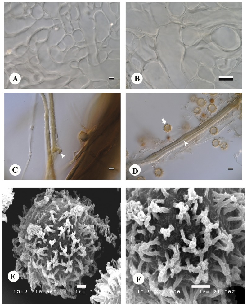
Figure 6. Astraeus sirindhorniae.
Exoperidium layers (A–B). (A) exoperidial subpellis, bar = 5 µm. (B) exoperidial subpellis (innermost), bar = 10 µm. (C) rhizomorph hyphae with clamp connection (arrowhead), bar = 5 µm. (D) capillitium hyphae displaying continuous lumen (arrowhead) and basidiospore (arrow), bar = 5 µm. (E–F) spore ornamentation demonstrated coalescent spines in groups, bar = 1 µm. A–D magnification at 1,000×.

