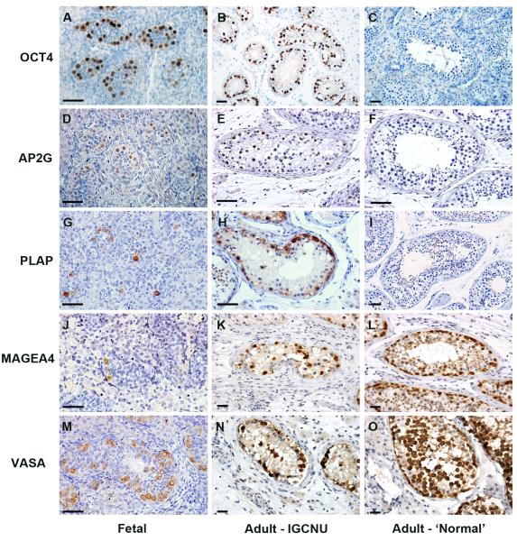
Figure 1.
Expression of gonocyte markers (OCT4, AP2γ and PLAP; A-I) and spermatogonial markers (MAGEA4 and VASA; J-O) in human fetal testis, intratubular germ cell neoplasia containing tubules (Adult – intratubular germ cell neoplasia) and tubules from adult testis with active spermatogenesis (Adult – ‘Normal’). Gonocyte proteins are detected in human fetal germ cells and intratubular germ cell neoplasia cells, but are absent from the germ cells in tubules with apparently normal spermatogenesis; whilst spermatogonial proteins are expressed in germ cells in all tissue types. Human fetal samples are 14 (A,D), 16 (J) and 18 weeks (G,M) gestation. Scale bar = 50μm.

