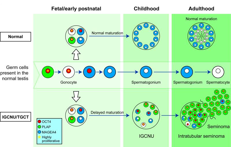
Figure 9.
Schematic for germ cell maturation and proliferation in germ cells during transition from gonocyte to intratubular germ cell neoplasia and testicular germ cell cancer (bottom). Germ cell maturation from gonocyte to initiation of spermatogenesis is represented in the testis during the different stages of life (middle). For comparison, germ cell differentiation in the normal testis is also shown (top). Germ cells in the fetal testis may exhibit delayed maturation with persistence of gonocyte markers through childhood. A variety of germ cell protein profiles are present in the intratubular germ cell neoplasia tubule, however it is the cells expressing exclusively gonocyte proteins (with no spermatogonial proteins) that are more proliferative and contribute to the majority of the cells in intratubular seminoma and subsequently invasive seminoma. Cells expressing spermatogonial proteins are occasionally seen in the tubule or resultant tumour but exhibit low proliferation rates. Expression of OCT4 (red), PLAP (green) and MAGEA4 (blue) is shown for individual cells and cells with high rates of proliferation are indicated (yellow asterisk)

