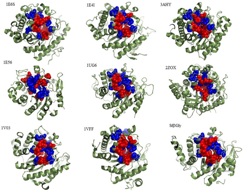
Figure 1. Structural comparison of β-glucosidases showing the active site residues (red) and sector A positions (blue).
Myrosinase from Sinapis alba (1E6S); β-glucosidase A from Paenibacillus polymyxa (1EI4); β-glucosidase from Trichoderma reesei (3AHY); β-glucosidase Zmglu from Zea mays (1E56); β-glucosidase from Thermus thermophilus (1UG6); Human cytosolic β-glucosidase (2ZOX); SbDhr from Sorghum bicolor (1V03); β-glucosidase from Pyrococcus horikoshii (1VFF); β-glucosidase from Spodoptera frugiperda Sfβgly. The distances between sector A and the active site residues are shorter than 4.5 Å. The structures were visualized using PyMOL software.

