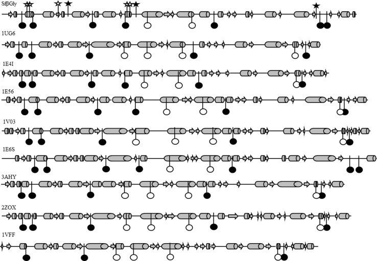
Figure 2. Distribution of sector S positions on the secondary structure of β-glucosidases.
β-glucosidase from Spodoptera frugiperda Sfβgly; β-glucosidase from Thermus thermophilus (1UG6); β-glucosidase A from Paenibacillus polymyxa (1E4I); β-glucosidase Zmglu from Zea mays (1E56); β-glucosidase SbDhr from Sorghum bicolor (1V03); myrosinase from Sinapis alba (1E6S); β-glucosidase from Trichoderma reesei (3AHY); Human cytosolic β-glucosidase (2ZOX); β-glucosidase from Pyrococcus horikoshii (1VFF). α-Helices are represented by cylinders, β-strands by arrows and loops by lines. Sector S positions are shown as circles, whereas non-sector S positions are shown as stars. The symbols (circles or stars) in black indicate positions placed at loops, whereas white symbols mark positions at helices or strands.

