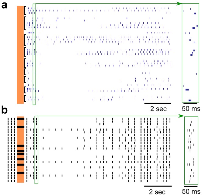
Figure 2. Post-stimulus responses of multiple neurons to brief epoch of high-frequency stimulation.
(a) Rasters of repeated trials across 9 different human thalamic neurons to 0.5 seconds of stimulation at 200 Hz and with stimulus intensities between 1.5–5 µA. (b) Single trial rasters across the 24 model neurons in response to 0.5 seconds of stimulation at 200 Hz and with a stimulus intensity of 1.5 V. The vertical shaded region indicates the time when stimulation was on. Insets to the right are expanded in the time axis to illustrate sample individual action potentials within burst episodes. The characteristic post-stimulus bursting followed by prolonged inhibition is present in both human and model neuron recordings. Note that the spontaneous bursting activity prior to stimulation is not shown in (a).

