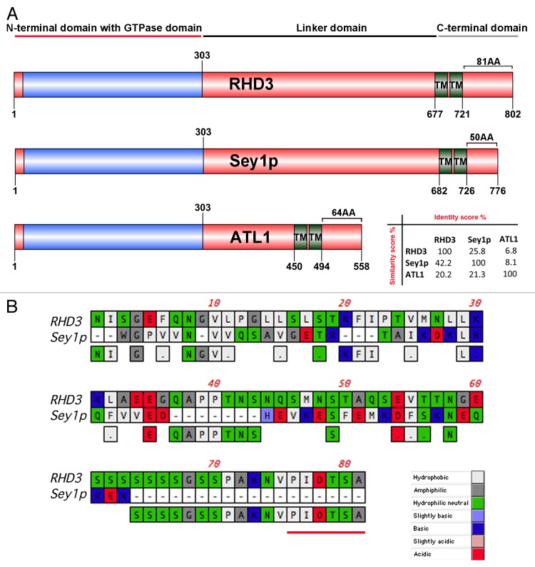
Figure 2. The overall structure of RHD3 and Sey1p is largely conserved although variations occur in the size of the CT extension. (A) Schematic representation of protein domains and motifs found in RHD3 (NCBI accession number: P93042), ATL1 (NCBI accession number: NP_056999) and Sey1p (NCBI accession number: NP_014808). Numbers indicate the amino acids. TM: transmembrane. Protein identity (%) vs. similarity (%) is indicated in the matrix. (B) Sequences of RHD3 (aa 722–802) and Sey1p (aa 727–776) were aligned using CLUSTALW and the GONNET matrix. Predicted PDZ binding motif in RHD3 is underlined in red.

An official website of the United States government
Here's how you know
Official websites use .gov
A
.gov website belongs to an official
government organization in the United States.
Secure .gov websites use HTTPS
A lock (
) or https:// means you've safely
connected to the .gov website. Share sensitive
information only on official, secure websites.
