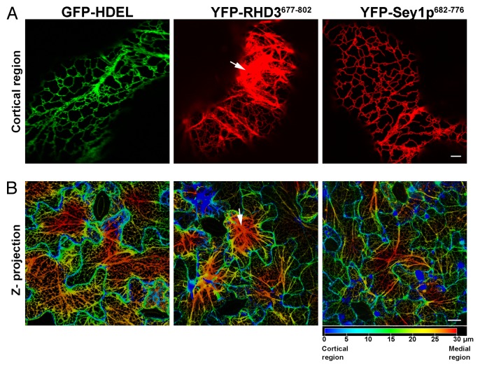
Figure 3. RHD3677–802 but not Sey1p682–776 alters ER structure. Confocal microscopy images of the ER in cells expressing either the ER marker GFP-HDEL, YFP-RHD3677–802 or YFP-Sey1p682–776, viewed as single plane for single cells (A) or in a 30 μm z-depth code (B, 0 μm-upper level = blue color; 30 μm-lower depth = red color). In A, the images show that differently from cells expressing GFP-HDEL or YFP-Sey1p682–776, expression of YFP-RHD3677–802 causes aberrant organization of the ER structure. Arrows in A and B show defects in the ER structures that are visible only in cells expressing YFP-RHD3677–802. Bars = 5 μm (A); 20 μm (B).

An official website of the United States government
Here's how you know
Official websites use .gov
A
.gov website belongs to an official
government organization in the United States.
Secure .gov websites use HTTPS
A lock (
) or https:// means you've safely
connected to the .gov website. Share sensitive
information only on official, secure websites.
