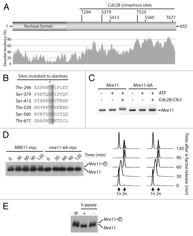
Figure 3. Mre11 is a substrate for phosphorylation by Cdc28. (A) Schematic representation of Mre11 and distribution of consensus Cdc28 Ser/Thr-Pro sites within the protein primary amino acid sequence. The predicted disorder of the protein (calculated by IUPred)65 is shown under the schematic. (B) Alignment of putative Cdc28 phosphorylation sites found in Mre11. The asterisk symbol indicates residues mutated to alanine. (C) Purified Mre11 and Mre11–6A proteins were phosphorylated in vitro using Cdc28-Clb2. Reaction mixtures were subsequently analyzed by SDS-PAGE and Coomassie staining. (D) Samples from MRE11-MYC and mre11–6A-MYC cultures were taken at indicated time points after release from a G1 block and subjected to SDS-PAGE analysis. DNA content determined by FACS shows that cell cycle progression was similar in wild-type and mre11–6A cultures (right). (E) Immunoprecipitated Mre11 was dephosphorylated with lambda phosphatase (or mock treated) prior to SDS-PAGE and immunoblot analysis.

An official website of the United States government
Here's how you know
Official websites use .gov
A
.gov website belongs to an official
government organization in the United States.
Secure .gov websites use HTTPS
A lock (
) or https:// means you've safely
connected to the .gov website. Share sensitive
information only on official, secure websites.
