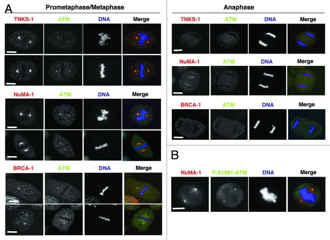
Figure 4. (A) Mitotic localization of ATM, BRCA1, NuMA1, and TNKS1. HeLa cells were fixed and processed for immunofluorescence and confocal microscopy analysis. BRCA1, NuMA1, and TNKS1 were stained in red, ATM in green, and DNA in blue. Mitotic phases are indicated. (B) HeLa cells were fixed and processed for immunofluorescence. NuMA1 was stained in red, P-Ser1981-ATM in green, and DNA in blue. Scale bars, 10 μm.

An official website of the United States government
Here's how you know
Official websites use .gov
A
.gov website belongs to an official
government organization in the United States.
Secure .gov websites use HTTPS
A lock (
) or https:// means you've safely
connected to the .gov website. Share sensitive
information only on official, secure websites.
