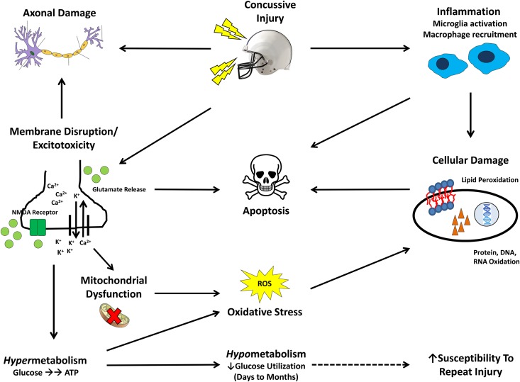
FIGURE 1.
Molecular cascade of events after a mild traumatic brain injury. The initial mechanical injury causes mild membrane disruption in the nerve, axonal damage, and indiscriminate neurotransmitter (glutamate) release and activation of ion channels, such as the NMDA receptor. Deregulation of Na+/K+/Ca2+ flux leads to excitotoxicity—a massive influx of calcium and an efflux of potassium and the release of the neurotransmitter glutamate. Ca2+ influx also exacerbates damage to the axonal structure and causes mitochondrial dysfunction. ATP-dependent Na/K pumps function at an elevated capacity, creating a hypermetabolic state generating oxidative stress that can result in cellular damage. Glucose stores become depleted due to the hypermetabolic state, resulting in a hypometabolic state, with low glucose utilization. This hypometabolic state may last for months in severe cases, and, during this time, the brain may be particularly vulnerable to repeated injury (dashed arrow). Concurrently, inflammation due to microglial activation occurs soon after the concussive injury, causing damage to cellular structures. Ultimately, the combination of oxidative stress, inflammation, excitotoxicity, mitochondrial dysfunction, and axonal damage results in neuronal apoptosis. The ω-3 FA DHA has been shown to address several of the hallmark pathologic features of this injury, such as excitotoxicity, oxidative stress, and inflammation. NMDA, N-methyl-d-aspartate; ROS, reactive oxygen species.

