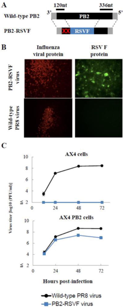
Fig. 2. Characterization of the PB2-RSVF virus.

(A) Schematic diagram of wild-type PB2 and PB2-RSVF vRNAs. PB2(120)FRSV(336) vRNA possesses the 3′ non-coding sequence of the PB2 vRNA, 120 nt of the 3′ coding sequence of the PB2 vRNA with two ATG mutations (red crosses), the full-length RSV F gene, 336 nt of the 5′ coding region of the PB2 vRNA, and the 5′ non-coding sequence of PB2 vRNA. The non-coding and coding sequences of the PB2 vRNA are represented by the gray and black bars, respectively. The RSV F gene is represented by the blue bar. (B) Expression of RSV F protein in PB2-RSVF virus-infected cells. AX4/PB2 cells were infected with wild-type PR8 (top panels) or PB2-RSVF (bottom panels) virus at an m.o.i. of 0.1. At 16 h postinfection, the cells were fixed and stained with anti-influenza virus NP (left panels) and anti-RSV F (right panels) antibodies. (C) Growth kinetics of the PB2-RSVF virus. AX4 and AX4/PB2 cells were infected with wild-type PR8 or PB2-RSVF virus at an m.o.i. of 0.001. Supernatants collected at the indicated time points were assayed for infectious virus by use of plaque assays in AX4/PB2 cells.
