
Figure 3. Stat3 and ERK1/2 signaling mediate the biological effects of IL-6 in induction of migratory PC cell phenotype.
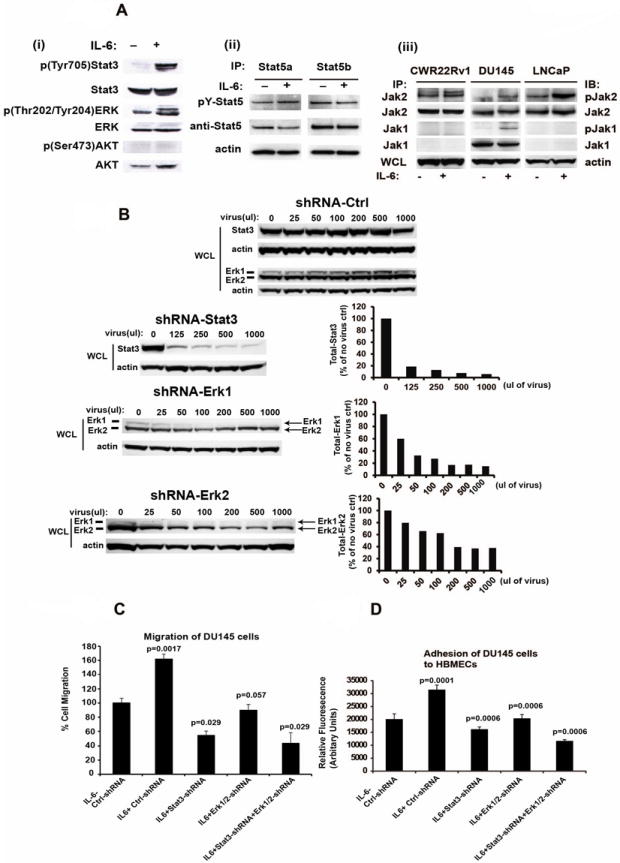
(A) IL-6 activates phosphorylation of Stat3, ERK1/2 and Jak1/2 but not Stat5 or Akt. (i) DU145 cells were serum-starved (0% FBS) overnight followed by stimulation of the cells with IL-6 (5 nM) for 20 min and immunoblotted for p(Tyr705)Stat3, Stat3, p(Thr202/Tyr204)ERK, ERK1/2 or p(Ser473)Akt or Akt. (ii) Stat5a and Stat5b were immunoprecipitated (IP) and immunoblotted for pYStat5a/b and total Stat5a/b. Whole cell lysates of parallel samples were immunoblotted for actin. (iii) CWR22Rv1, DU145 and LNCaP cells were serum-starved overnight followed by stimulation with IL-6 (5 nM) for 20 min. Jak1 and Jak2 were immunoprecipitated and blotted with anti-phosphotyrosine (4G10), anti-Jak1 or anti-Jak2 mAbs. (B) Lentivirus expressing shRNAs targeting Stat3, ERK1 and ERK2 expression in DU145 cells. DU145 cells infected with each lentiviral construct were immunoblotted with anti-Stat3, anti-actin, anti-ERK1 or anti-ERK2 antibodies. (C) Inhibition of Stat3 and/or ERK1/2 by RNA interference blocks IL-6-induced PC cell migration. DU145 cells were cultured in the presence of 1% CS-FBS and infected with lentiviruses expressing control-shRNA, Stat3-shRNA and/or ERK1/2-shRNA (MOI=5) and treated with or without IL-6 (5 nM) for 72 h. Cells were counted and equal numbers of cells in each treatment group were assayed for migration in Boyden chambers for 16 h. (D) Inhibition of Stat3 and/or ERK1/2 signaling blocks IL-6-induced increase in adhesion of PC cells to endothelial cells. DU145 cells were cultured in the presence of 1% CS-FBS and infected with lentiviruses expressing control-shRNA, Stat3-shRNA and/or ERK1/2-shRNA (MOI=5) and treated with or without IL-6 (5 nM) for 72 h. Cells were stained with a fluorescent dye and allowed to adhere to HBMEC cells for 60 min followed by quantitation of adhered cells by fluorescent reader. Averages of three independent experiments are shown.