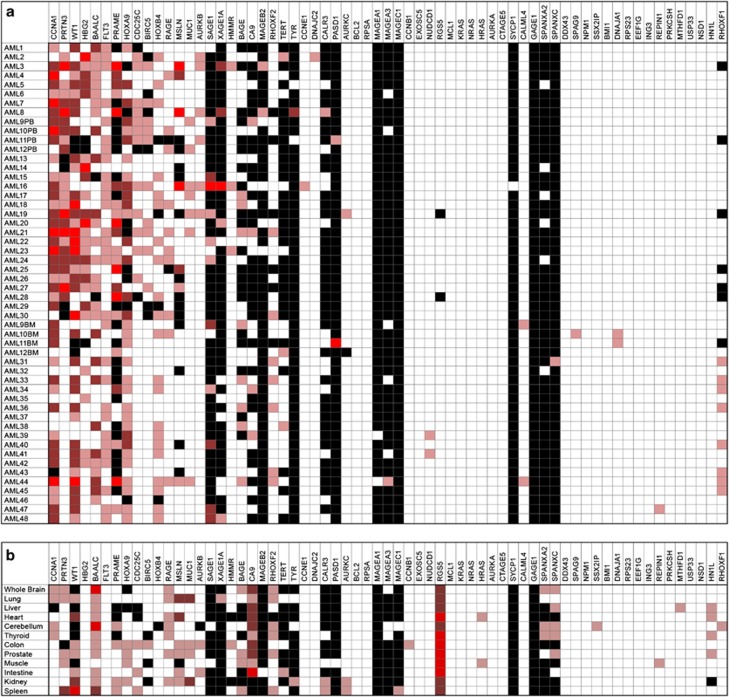
Figure 1.
Expression of proposed leukemia associated antigens in acute myeloid leukemia (AML) patient samples and healthy tissues. (a) No single antigen was expressed in all cases of AML and many proposed antigen candidates are not frequently overexpressed in AML. BM, bone marrow; PB, peripheral blood. Fold change OE compared with median expression in healthy donors where light red indicates OE of 5–50 × , red indicates OE of 50–500 × , bright red indicates OE >500 × . Black indicates no detectable expression; white indicates expression values seen in similar range as healthy donors. First 30 AML samples listed were from PB and are therefore compared with healthy donor PB, the remaining 18 are from BM and are compared with expression in healthy donor BM. (b) Antigen expression in various human tissue types. Compared with median expression in healthy donors using same heat-map schema same as above.

