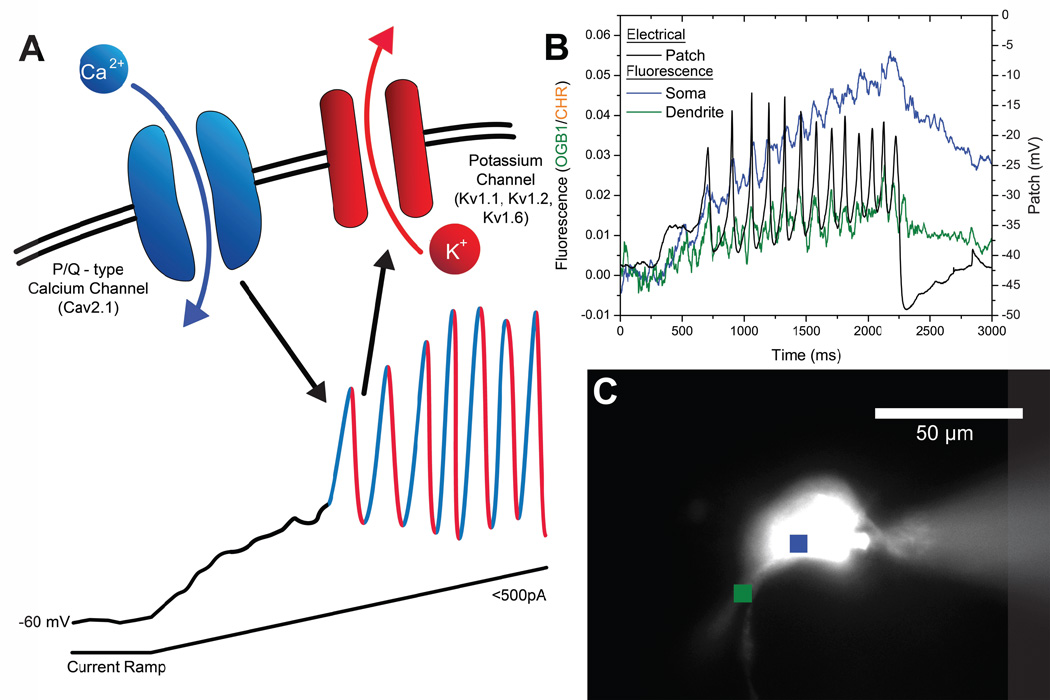
Figure 1. Model of high threshold, voltage-dependent P/Q-type calcium channel oscillations, and visualization of calcium transients in the dendrite of a PPN neuron.
A) Application of a ramp stimulus in the presence of synaptic blockers and TTX slowly depolarizes the membrane, avoiding activation of potassium channels. Once the voltage-dependent high threshold of calcium channels is reached, ~ −30 mV, membrane oscillations are observed. Addition of the specific P/Q-type calcium channel blocker ω-agatoxin-IVA will eliminate the oscillations, demonstrating that the depolarizing phase is due to P/Q-type calcium channels (Cav2.1). Addition of the delayed rectifier-like potassium channel blocker dendrotoxin also blocks the oscillations, demonstrating that the repolarizing phase is due to these potassium channels (Kv1.1, Kv1.2, Kv1.6) (Kezunovic et al. 2012). B) Electrophysiological recording of a PPN cell during ramp-induced membrane oscillations (black record), while region of interest calcium transients were imaged from the cell body (blue record) and one of the dendrites (green record). The peaks of the calcium oscillations coincided with the peaks in the electrical recording, and different dendrites (not shown) manifested calcium transients coinciding with different of the peaks of the soma recording (Hyde et al 2013b). C) Locations of the regions of interest shown in B in a photomicrograph of a PPN cell injected with fluorescent dye, with the microelectrode evident on the right.

