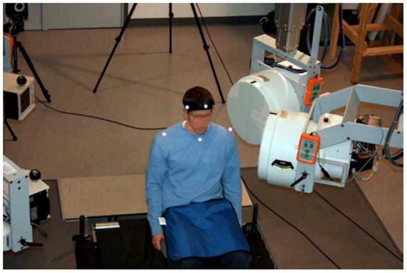
Figure 2.

Biplane x-ray data collection system. X-ray tubes (left) directed X-rays through the subject to image intensifiers (right). 2.5 ms X-ray pulses (70 kV, 160 mA) were generated by cardiac cine-angiography generators at a rate of 30 Hz and images were collected by high-speed cameras synchronized to the x-ray pulses.
