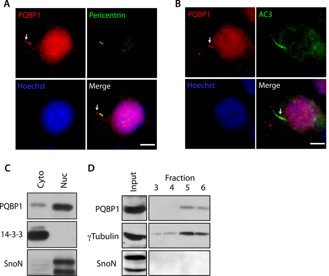
Figure 3. PQBP1 localizes at the base of the cilium and centrosome.
(A and B) Hippocampal neurons were subjected to immunocytochemistry with the PQBP1 antibody together with the Pericentrin antibody (A), or AC3 antibody (B). Arrows indicate PQBP1 immunoreactivity at the cilium. Scale bar = 5 µm.
(C) Cytoplasmic and nuclear fractions isolated from rat cortical neurons were immunoblotted using the PQBP1, 14-3-3 and SnoN antibodies. 14-3-3 and SnoN served as the cytoplasmic and nuclear marker, respectively.
(D) Centrosomal fractions prepared from rat cortical neuron lysates were immunoblotted using the PQBP1, γTubulin, and SnoN antibodies. PQBP1 and γTubulin cofractionated, suggesting that PQBP1 is present at centrosome. SnoN served as negative control.
See also Figure S3

