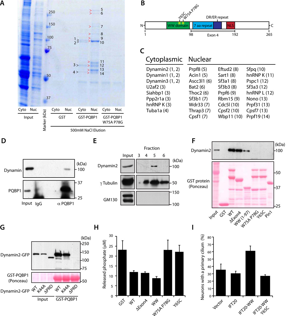
Figure 4. PQBP1 forms a complex with Dynamin 2 and thereby inhibits the GTPase activity of Dynamin 2.
(A) Proteins coprecipitated with GST-PQBP1, GST-PQBP1 W75A P78G, or GST were analyzed by SDS-PAGE and Coomassie Brilliant Blue staining. Numbers and arrowheads indicate bands analyzed by mass spectrometry.
(B) Schematic of PQBP1 protein domain structure. The WW domain, 7 aa repeat, DR/ER repeat, and nuclear localization signal (NLS) are indicated.
(C) Identity of PQBP1-associated proteins in cytoplasmic and nuclear fractions of cortical neuron lysates. Numbers in parenthesis indicate protein band in (A).
(D) Cytoplasmic fraction of P6 rat brain lysate was immunoprecipitated with the PQBP1 antibody or rabbit IgG and immunoblotted with the Dynamin or PQBP1 antibody. Asterisk denotes a non-specific band.
(E) Centrosomal fractions isolated from rat cortical neurons were immunoblotted using the Dynamin 2, γTubulin, and GM130 antibodies. Dynamin 2 and γTubulin cofractionated, suggesting Dynamin 2 is present at the centrosome. GM130 served as a negative control.
(F) Lysates of rat cortical neurons subjected to pull-down assay with GST, GST-PQBP1, GST-PQBP1 ΔExon4 (Δ98–192), GST-PQBP1 WW (1-97), GST-PQBP1 W75A P78G, GST-PQBP1 Y65C or GST-Pin1, and immunoblotted with Dynamin 2 antibody or stained with Ponceau S.
(G) Lysates of 293T cells transfected with Dynamin 2-GFP, Dynamin 2 K44A-GFP or Dynamin 2 ΔPRD-GFP were subjected to a pull-down assay with GST-PQBP1 and immunoblotted with the GFP antibody or stained with Ponceau S.
(H) Phosphate released from GTP by Dynamin 2 was measured in the presence of GST, GST-PQBP1, GST-PQBP1 ΔExon4 (Δ98–192), GST-PQBP1 WW (1-97), GST-PQBP1 W75AP78G, or GST-PQBP1 Y65C.
(I) The percentage of hippocampal neurons bearing a primary cilium was significantly higher at DIV3 in neurons transfected with GFP-IFT20-WW compared to control vector, GFP-IFT20 or GFP-IFT20-WW Y65C (p < 0.01; ANOVA). Total of 387 neurons were measured. See also Figure S4

