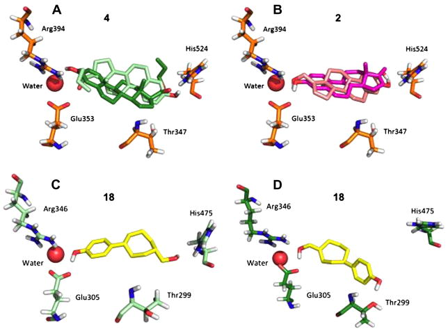
Figure 2.
Lowest energy docking poses from clusters where ligands were predicted to bind in two modes (A–B). The human ERα estrogen receptor that was used was in the agonist conformation (PDB code 1ere; chain A). Panel C shows the predicted binding orientation for 18 in ERβ, agonist conformation (PDB code 2jj3; chain A). Panel D shows the predicted binding orientation for 18 in ERβ, antagonist conformation (PDB code 1l2j; chain A).

