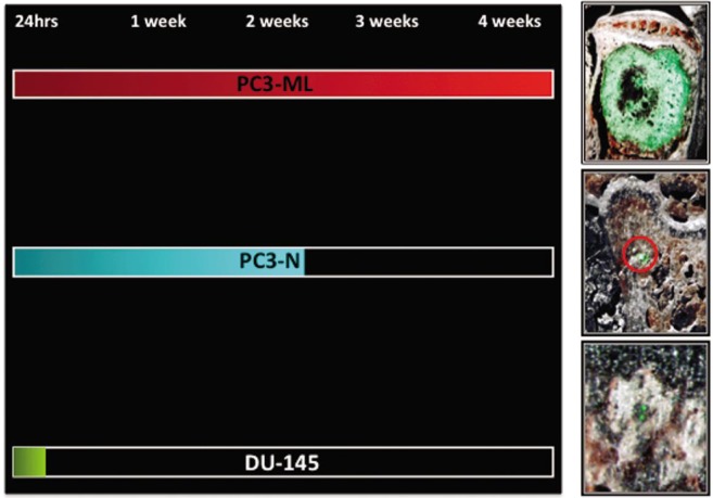
Figure 1. Survival and progression at the skeletal level of prostate cancer cell types expressing different levels of PDGFRα.
The PC3-ML sub-line expressed higher levels of the receptor and produced macroscopic skeletal metastases in mice inoculated with cancer cells in the hematogenous circulation via the left cardiac ventricle injection. PC3-N cells expressed lower levels of PDGFRα than did PC3-ML cells and could only survive two weeks in the bone after their dissemination. DU-145 cells were found negative to PDGFRα expression and disappeared from the skeleton between 72 h and one week post inoculation.

