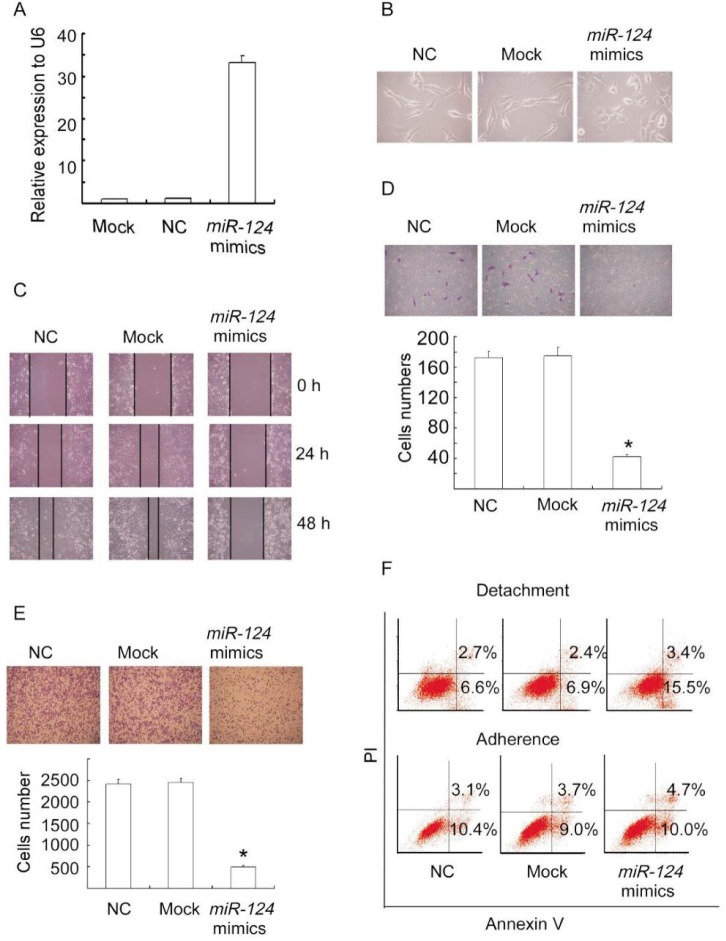
Figure 2. miR-124 expression suppresses metastasis-relevant traits in vitro.
A, overexpression of miR-124 in MDA-MB-231 cells was detected by quantitative real-time PCR (qRT-PCR). MDA-MB-231 cells were transfected with miR-124 mimics, negative control (NC), or mock for 48 h and the RNAs were extracted. B, the morphology of MDA-MB-231 cells changed from spindle-shaped to round as observed under a microscope after transfection with miR-124 mimics, negative control, or mock for 48 h. C, the wound-healing assay shows decreased cell motilities in miR-124 ectopically expressed MDA-MB-231 cells. MDA-MB-231 cells transfected with miR-124 mimics, negative control, or mock were cultured with serum-free medium for 24 h and scraped to created acellular area. The spread of wound closure was observed 24 h and 48 h after scrape and photographed under a microscope. D, ectopic expression of miR-124 suppresses the migratory capacity of MDA-MB-231 cells. MDA-MB-231 cells transfected with miR-124 mimics, negative control, or mock for 48 h were placed in serum-free medium and added to the upper chamber of transwell plates. Medium containing 10% serum was added to the lower chamber as a chemoattractant. The migratory capacity was assessed by calculating the filtered cells. Columns, mean of three independent experiments; bars, SD. *P < 0.01, vs. negative control and mock cells. E, ectopic expression of miR-124 reduces the adhesion of MDA-MB-231 cells to fibronectin. MDA-MB-231 cells transfected with miR-124 mimics, negative control, or mock for 48 h were detached from culture dishes with trypsin and suspended in serum-free medium. The suspended cells were seeded to 24-well plates coated with fibronectin for 30 min and then observed under a microscope. Columns, mean of three independent experiments; bars, SD. *P < 0.01, vs. control and mock cells. F, ectopic expression of miR-124 increases the sensitivity of MDA-MB-231 cells to anoikis. MDA-MB-231 cells transfected with miR-124 mimics, negative control, or mock for 24 h were detached from culture dishes with trypsin and seeded to anoikis plates for another 72 h. Apoptotic cells were evaluated by staining with FITC-annexin V and propidium iodide (PI) and analyzed with FACS.

