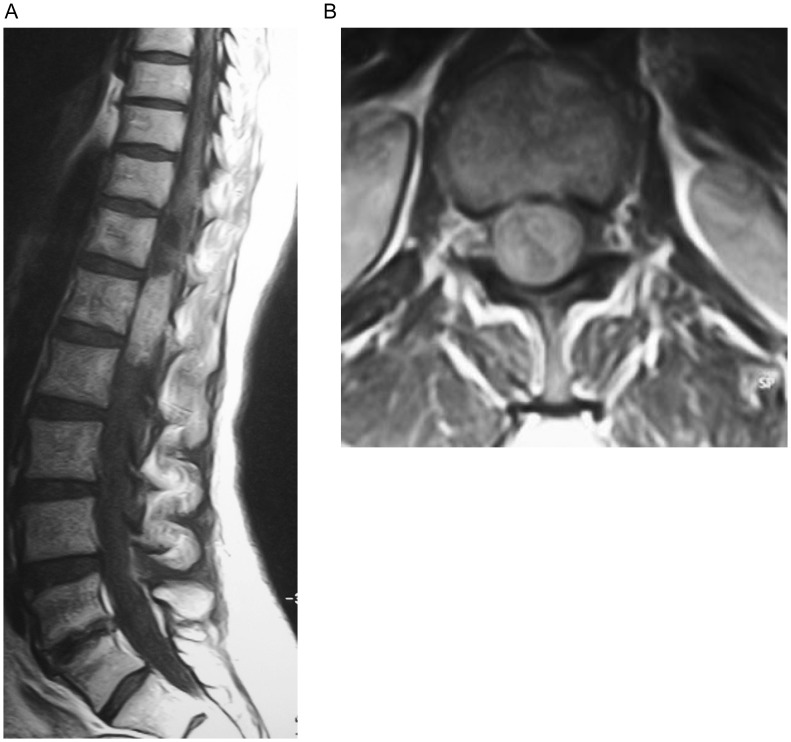
Figure 1. Magnetic resonance (MR) images of schwannoma of the conus medullaris in a 49-year-old woman with chronic low back pain and sciatica.
A, the sagittal T1-weighted MR image shows an intradural, isointense lesion of the conus medullaris. B, on the axial T1-weighted MR images, this lesion was heterogeneously enhanced with contrast. The spinal nerves are displaced laterally.

