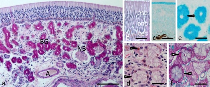
Fig. 1.
Histological features of the sheep olfactory mucosa. (a) Transverse section of the olfactory mucosa stained with Periodic Acid Schiff (PAS). Bar=100 µm. Higher magnification of the olfactory epithelium stained with (b) PAS and (c) alcian blue (pH 2.5). Higher magnification of the Bowman’s glandular acini stained with Hematoxylin and Eosin (d), alcian blue (pH 2.5) (e) or Crossmon’s trichrome (f). Arrowheads indicate Bowman’s glandular acinar cells. A, artery; BG, Bowman’s glands; NB, nerve bundle; OBC, olfactory basal cells; ORC, olfactory receptor cells; OSC, olfactory supporting cells. Bar=30 µm in (b)–(f).

