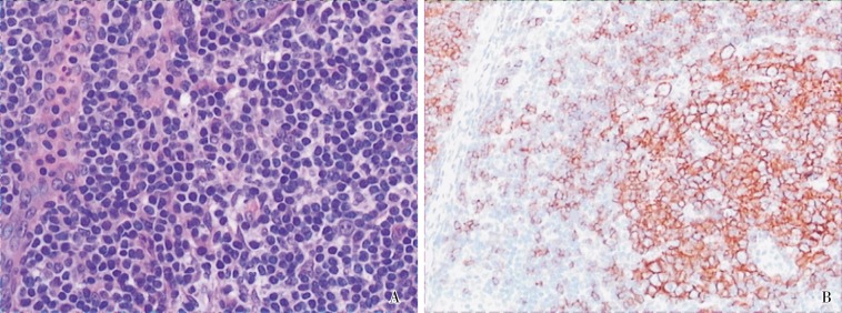
Figure 2. Histological examination of the thoracic tumor from the 55-year-old woman. The patient underwent thoracic operation in April 2009 at another hospital, but the mass could not be completely resected. Therefore, a biopsy was performed for histological examination. A, lymphocytes diffuse in the mass and vessels penetrate into the follicles, indicating lymphoid hyperplasia (HE ×20). B, positive staining for CD20 shows on the cell membrane (IHC × 10).

