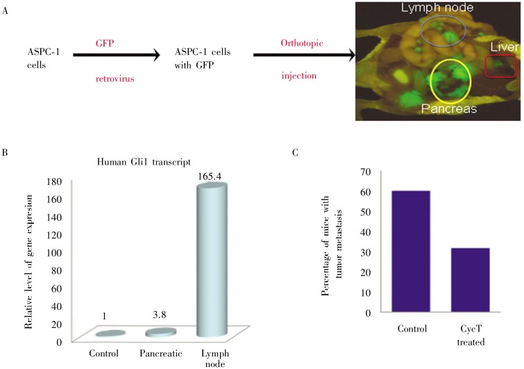
Figure 7. Effects of CycT on lymph node metastasis of pancreatic cancer.
A shows the orthotopic model of pancreatic cancer metastasis. Mice were killed 6 weeks after GFP-expressing ASPC-1 cells were injected into the pancreas of NGS mice. GFP-positive tissues indicate tumor and its metastases in different organs (noticeably lymph nodes and liver). B shows expression of Hh target gene GM, indicating activation of hedgehog signaling pathway during pancreatic cancer metastasis. C shows the percentage of mice with lymph node metastases of tumor in two groups (control or CycT-treated), with 10 mice in each group.

