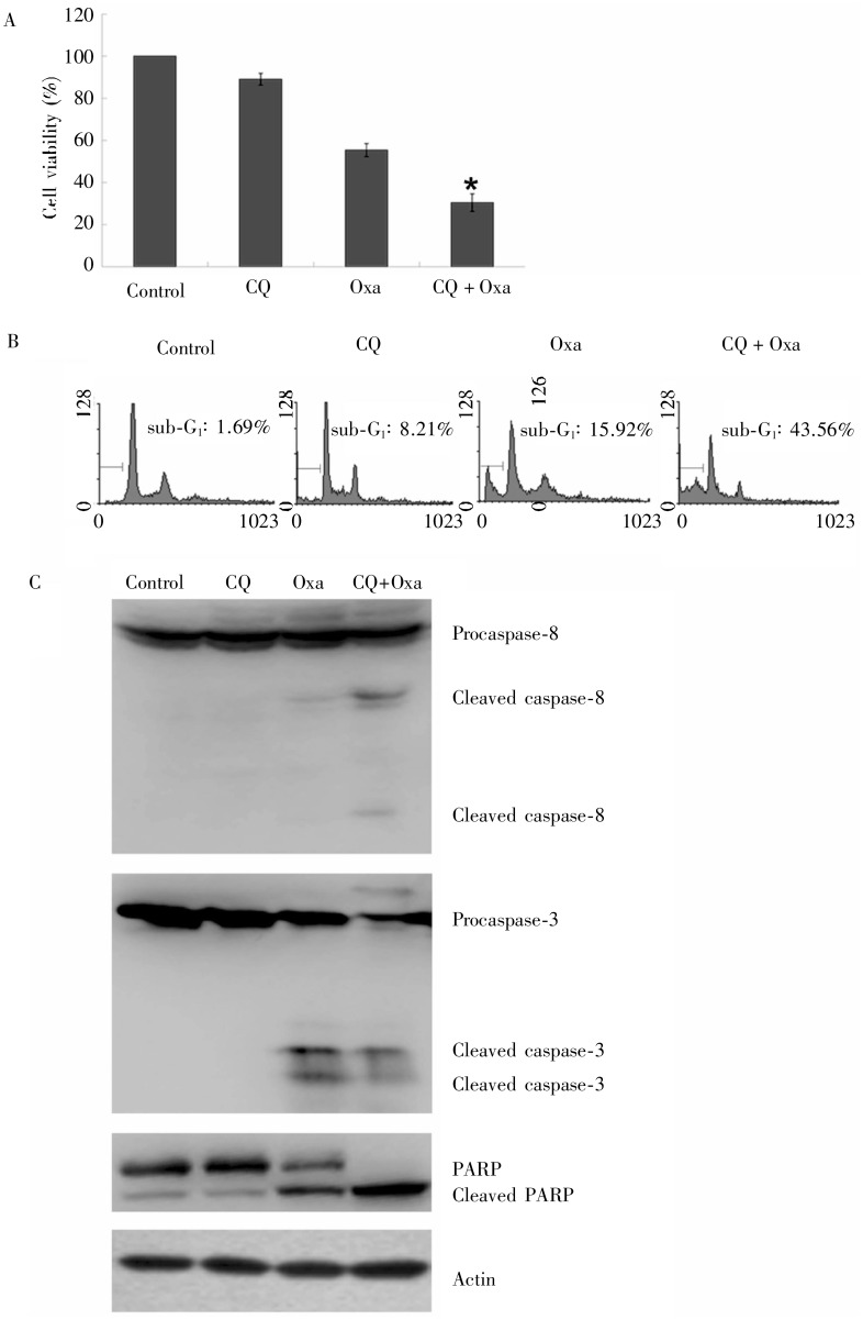
Figure 4. The effect of oxaliplatin and chloroquine on survival and apoptosis in gastric cancer MGC803 cells.
MGC803 cells were exposed to 20 µg/mL oxaliplatin and 20 µmol/L chloroquine for 24 h. A, cell survival was determined by MTT assay. *Compared to oxaliplatin alone, the combination of oxaliplatin and chloroquine significantly inhibited cell survival (P < 0.05). B, cell apoptosis was quantified with flow Cytometry. Compared to oxaliplatin alone, the combination of oxaliplatin and chloroquine significantly enhanced cell apoptosis. C, the cleavage of caspase-8, caspase-3, and PARP protein was detected by Western blotting, indicating the increase of cell apoptosis. CQ, chloroquire; Oxa, oxaliplatin.

