
Figure 2.
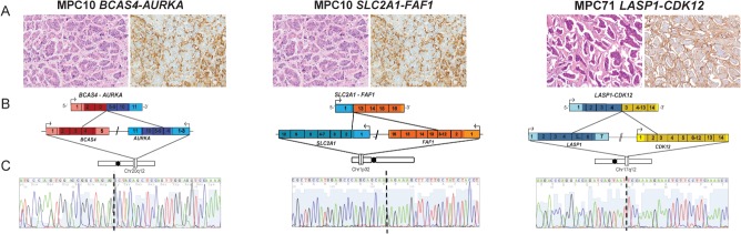
Structure of validated high-confidence fusion genes in micropapillary carcinomas. Sanger sequencing validation of functionally assessed fusion genes in the index cases of micropapillary carcinomas (MPCs) (BCAS4–AURKA and SLC2A1–FAF1 in MPC10 and LASP1-CDK12 in MPC71). (A) Haematoxylin and eosin (H&E) and epithelial membrane antigen (EMA) staining of representative areas of the MPCs harbouring the BCAS4–AURKA, SLC2A1–FAF1, and LASP1–CDK12 fusion genes (10× original magnification). (B) Schematic representation of nominated fusion transcripts. Fusion junctions with respective exon numbers are shown, while paler colours indicate 3′ and 5′ UTRs. (C) cDNA level sequence chromatograms spanning the junction (dotted line) of the fusion transcript.