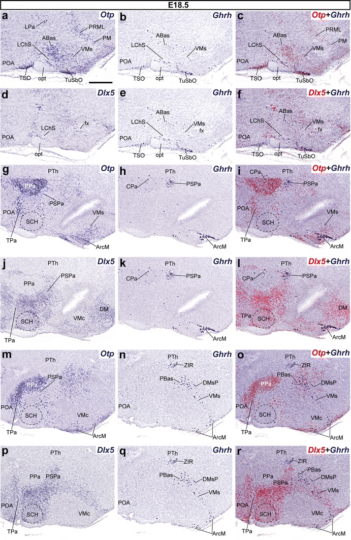
Fig. 11.

Series of parasagittal sections through an E18.5 embryo from lateral (a–f) to medial (g–r) levels, correlating the expression pattern of the indicated reference markers (Otp, Dlx5) with the presence of Ghrh+ cells. Corresponding digital overlaps with pseudocolored reference markers and Ghrh signals are marked with a plus symbol. Scale bar 300 μm
