Fig. 12.

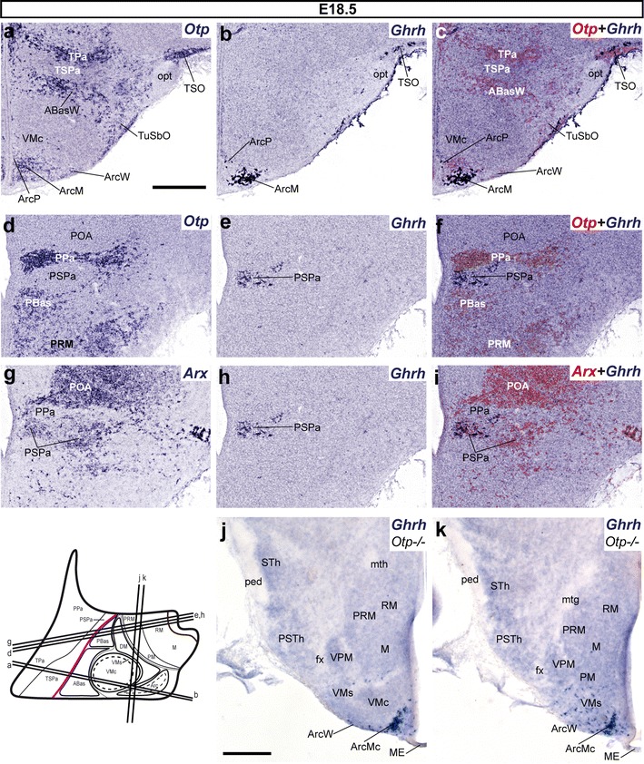
Ghrh+ cells mapped at E18.5. a–c, d–i Alternate transverse sections at two different rostrocaudal levels, correlating the expression patterns of indicated reference markers (Otp, Arx) with the presence of Ghrh+ cells. Corresponding digital overlaps of pseudocolored reference markers and Ghrh signal are marked with a plus symbol. j, k Horizontal sections at two different dorsoventral levels of Otp-null E18.5 embryos showing basal Ghrh+ cells largely restricted to the Arc tuberal area within THy (ArcM, ArcW), but extending as well into the nearby ventral shell of the VM nucleus (VMs). Transverse and horizontal section planes are shown in the adjacent schematic insert. mth mamillothalamic tract, ped cerebral peduncle,STh subthalamic nucleus, PSTh parasubthalamic nucleus, VPM ventral premamillary nucleus. Scale bar 300 μm
