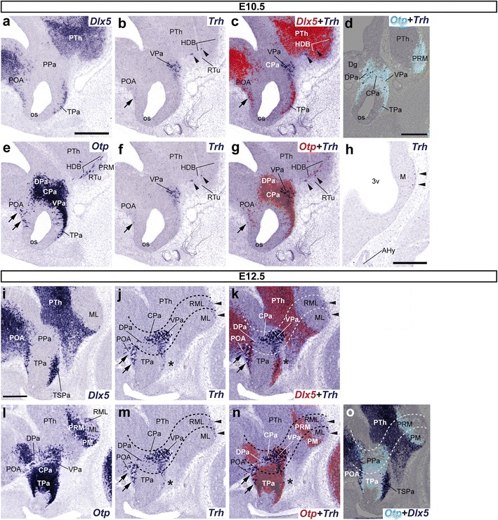
Fig. 3.

Sets of adjacent parasagittal sections from E10.5 (a–h) and E12.5 (i–o) embryos taken at lateral (a–g, i–o) and paramedian (h) levels correlate the presence of Trh+ cells with the indicated reference markers (Otp, Dlx5). Corresponding digital overlaps with pseudocolored reference genes and Trh signal are marked with a plus symbol. Transverse PHy (caudal) and THy (rostral) boundaries are indicated with dashed lines in j, k, m–o. Note that some Trh+ cells present at the surface of the mamillary/retromamillary region (arrowheads in h, j, k, m, n), as well as others intermixed with apparently migrated Otp+ cells within the extensive Dlx5 + POA, dorsal to the preopto-hypothalamic boundary (arrows in b, c, e–g, j, k, m, n). Few dispersed Trh+ cells are found in close proximity to the imprecise Otp/Dlx5 (TPa/TSPa) interface (asterisks in j, k, m, n) and close to the hypothalamo-diencephalic boundary (arrowheads in b, c, f, g). Scale bars 300 μm
