
Fig. 5.
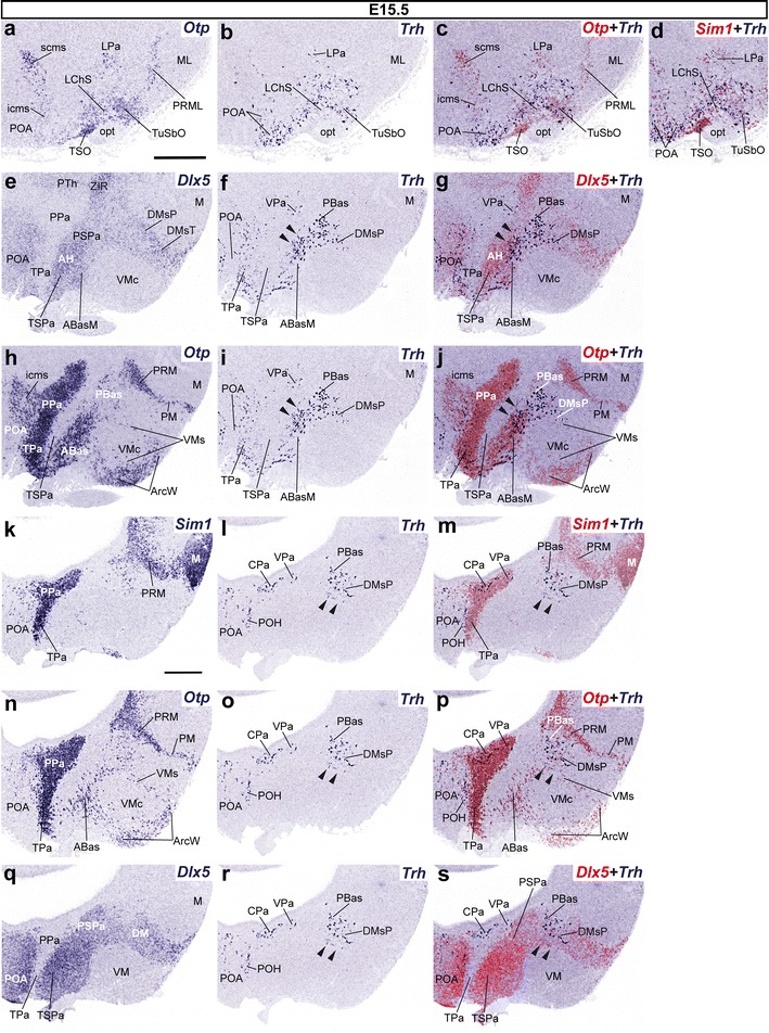
Parasagittal sections from an E15.5 embryo taken at lateral (a–d) and medial (e–s) levels, comparing in each case the expression pattern of the indicated reference genes (Otp, Sim1, Dlx5) with Trh+ cells. Corresponding digital overlaps with pseudocolored reference markers and Trh signal are marked with a plus symbol. Note that dispersed Trh+ cells lie within the borders of the Otp+/Dlx5 + PBas territory (arrowheads in f, g, i, j), as well as within the shell of the ventromedial nucleus (VMs; arrowheads in l, m, o, p, r, s). Scale bars 300 μm