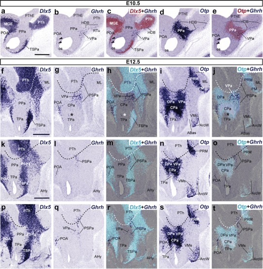
Fig. 8.

Lateral parasagittal sections from E10.5 (a–e) and E12.5 (f–t) embryos, correlating the presence of Ghrh+ cells with the indicated reference markers (Otp, Dlx5). Corresponding digital overlaps with pseudocolored reference markers and Ghrh cells are marked with a plus symbol. The transverse hypothalamo-diencephalic boundary is indicated with dashed lines in f–t. Arrowheads in b, c, e mark the presence of some dispersed Ghrh+ cells within the Dlx5+preoptic area, lying immediately dorsal to the preopto-hypothalamic limit and coinciding also with Otp-positive cells. Asterisks in g, h, j indicate some Ghrh+ cells in TPa. Scale bars 300 μm
