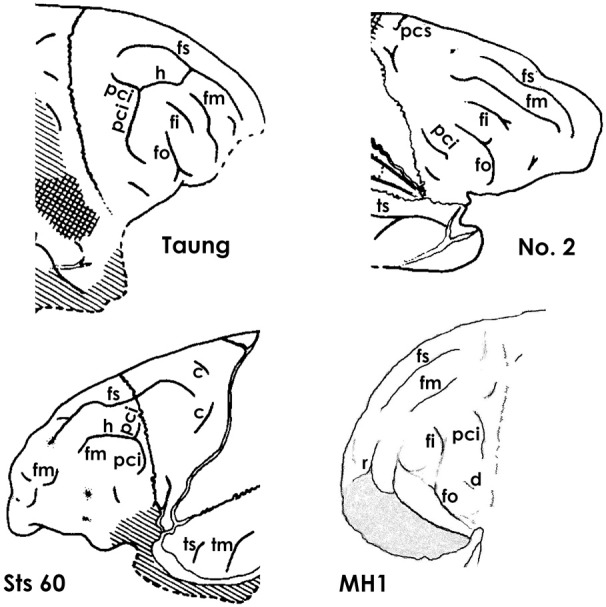
Figure 5.

The superior and middle frontal sulcus in Australopithecus endocasts. The middle frontal sulcus (fm) of MH1 (Australopithecus sediba) is identified here because (1) of its relationship to r (not identified by Carlson et al., 2011), (2) it does not appear to have been derived from (or proximal to) pci (which fi always is, Connolly, 1950), and (3) it courses approximately in the middle of the prefrontal cortex (hence its name). Taung, No. 2, and Sts 60 are endocasts from Australopithecus africanus. Note that all four australopithecines have separate branches of fm, which are rare in ape brains but typical of human brains, and that, in all four, fm is lateral to a long superior frontal sulcus (fs). Identifications of sulci: tm, middle temporal; ts, superior temporal; see legend to Figure 4 for other sulcal abbreviations. All illustrations except for MH1 are reproduced from Falk (1980). The identification of fi in Taung and No. 2 departs from Falk's (1980, 2009) earlier identification of that sulcus as r, but agrees with those of Dart (1929) and Schepers (1946). The line drawing for MH1 is based on the unlabeled photograph of the MH1 endocast from Carlson et al. (2011), reproduced on the left side of Figure 6. Compare the identifications for MH1 provided here with those of Carlson et al. reproduced on the right side of Figure 6.
