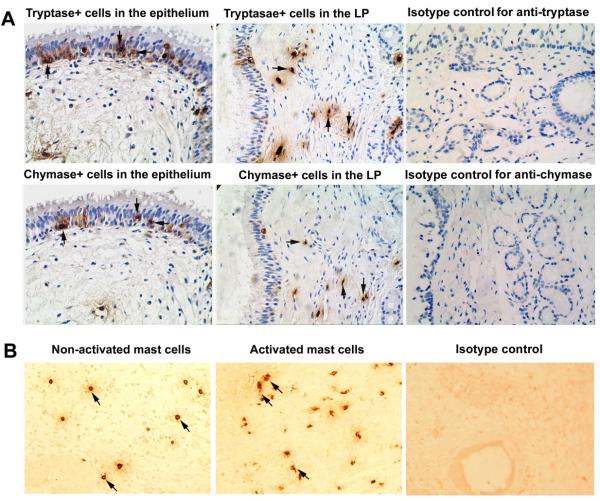
FIG 3.
A, Representative photomicrographs of tryptase and chymase immunohistochemical staining of consecutive sections of polyp tissues. Arrows in the same direction indicate the same cells in consecutive serial sections that are positive for both tryptase and chymase and defined as tryptase- and chymase-positive mast cells (MCTC). Tryptase-positive mast cells generally reflect the total population of mast cells (MCTot). LP, lamina propria. B, Representative photomicrographs of tryptase immunohistochemical staining of polyp tissue section showing activated and undegranulated mast cells. Degranulated mast cells were identified as cells that have discrete and relatively focal staining, but do not have well-circumscribed staining. In addition, extracellular staining is often observed in the area surrounding activated cells. The slides for observing activated mast cells were not counterstained with hematoxylin. Representative photomicrographs of immunohistochemical staining with respective isotype controls are also provided. Original magnification × 400.

