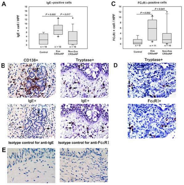
FIG 5.

IgE- and FcεRI-positive cells in tissues. A, The number of IgE-positive cells is increased in eosinophilic chronic rhinosinusitis with nasal polyps (CRSwNP). B, IgE-positive cells are mast cells and plasma cells. Representative immunostaining of consecutive sections from an eosinophilic CRSwNP subject with anti-IgE and anti-CD138 antibody, respectively; or with anti-IgE and anti-tryptase antibody, respectively. C, The number of FcεRI-positive cells is increased in eosinophilic CRSwNP. D, FcεRI expressing cells are mast cells. Representative immunostaining of consecutive sections from an eosinophilic CRSwNP subject with anti-FcεRI and anti-tryptase antibody, respectively. E, Representative photomicrographs of immunohistochemical staining with isotype controls for anti-IgE and anti-FcεRI antibody. In (B) and (D), arrows with the same direction indicate same cells in consecutive serial sections, original magnification × 400. HPF, high-powered field; Eos CRSwNP, eosinophilic CRSwNP; Non-Eos CRSwNP, non-eosinophilic CRSwNP.
