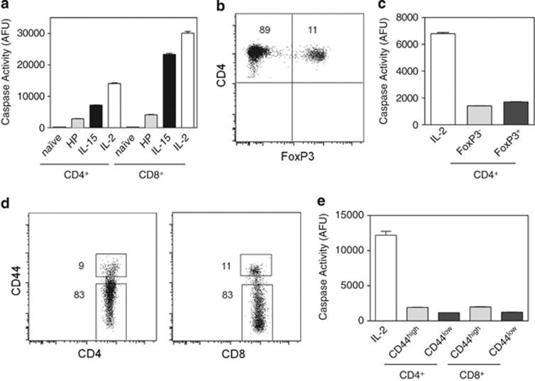
Figure 5.
Low levels of caspase activity in homeostatically proliferating, regulatory, naive, and memory T cells. (a) Caspase activity of T cells undergoing homeostatic expansion (HP) was compared with day 5 IL-2- or IL-15-cultured T cells and freshly isolated naive (CD44low) T cells. Homeostatically proliferating T cells were generated through adoptive transfer of wild-type lymphocytes into Rag1−/− mice and on day 14, donor CD4+ and CD8+ T cells were purified from Rag1−/− mice by cell sorting. (b and c) Freshly isolated CD4+ T cells from FoxP3GFP mice were sorted for FoxP3− and FoxP3+ cells and their caspase activity was compared with day 5 IL-2-cultured T cells. Numbers in flow cytometry quadrants indicate the percent positive cells. (d and e) Freshly isolated T cells from wild-type mice were sorted for CD4+CD44high, CD4+CD44low, CD8+CD44high, and CD8+CD44low cells and their caspase activity was compared with day 5 IL-2-cultured T cells

