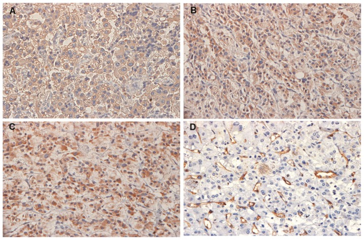
Figure 1.
Representative immunohistochemical detection of vascular endothelial growth factor (VEGF-A), vascular endothelial growth factor receptor 1 and 2 (VEGFR-1 and VEGFR-2) and microvessels in tumor cells from PHEO samples (all images were acquired using a magnification of 400×). (A) VEGF-A in a malignant tumor sample; (B) VEGFR-1 and (C) VEGFR-2 in a sample of sporadic tumor; and (D) CD31-positively stained microvessels in a sample of MEN 2B-associated PHEO.

