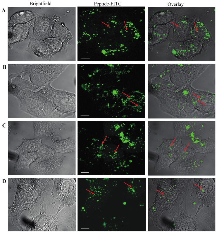
Figure 2.
Cellular distribution of SRC-1 bioportides. (A) TP10-SRC1LXXLL; (B) TP10-SRC11222–1245; (C) R7-SRC1LXXLL; (D) R7-SRC11222–1245. Confocal microscopy of MCF-7 cells exposed to 5 μM FITC-labeled peptides for 1 h. The cells were washed 3 times with stimulation medium before confocal microscopy of live cells was carried out. Nuclear localization of peptides is indicated with red arrows (63× magnification; scale bar 10 μm).

