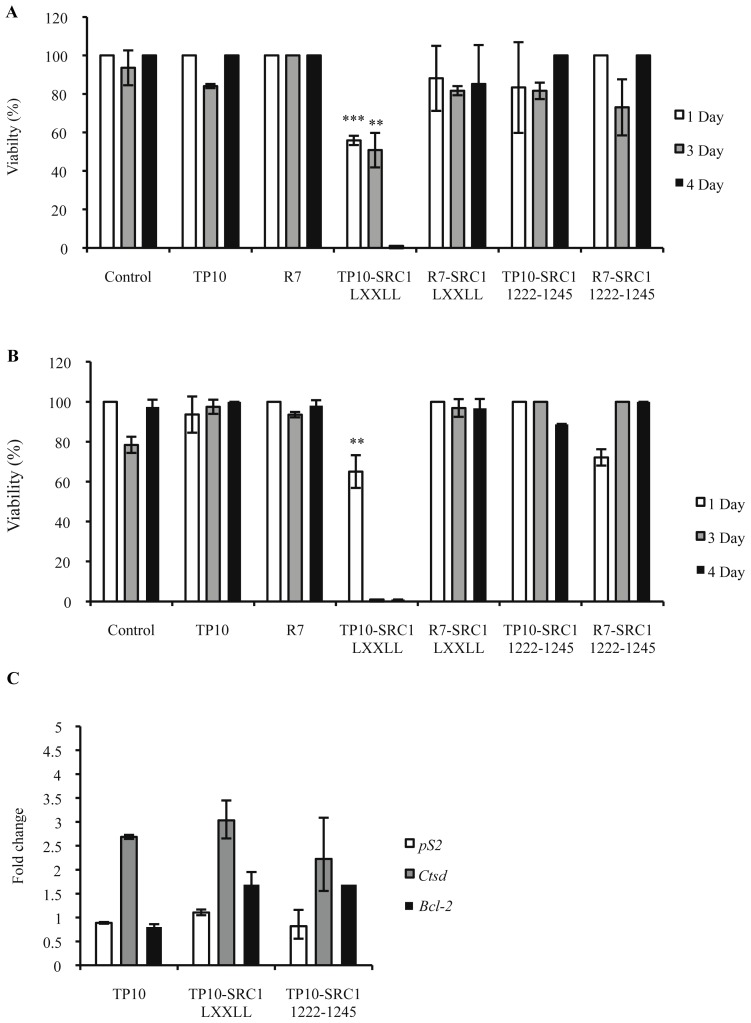
Figure 3.
Inhibition of cell viability by SRC-1 bioportides and their influence on ERα-mediated gene transactivation. (A) MCF-7 cells; (B) MDA-MB-231 cells. Cells were seeded in 24-well plates at low density and were treated every 24 h for 4 days with 5 μM peptide. Cell proliferation was measured after 1, 3, and 4 days of peptide treatment via a propidium iodide viability assay. Differences were found to be statistically significant with ** p < 0.01, *** p < 0.001; (C) Endogenous ERα target genes pS2, CTSD, and BCL2 mRNA expression analysis. MCF-7 cells were cultured with 5 μM of the indicated peptides and analyzed on day 2 of treatment. The fold change is the value compared with untreated cells. The values shown represent the mean values of one representative experiment, carried out in triplicate (mean ± SD). In total, three independent experiments were performed.

